Bronchoscopy
Flexible Bronchoscopy
Bronchoscopy is a procedure that lets doctors look inside air passages. It’s performed by a pulmonologist. During bronchoscopy, a thin tube (bronchoscope) is passed through your nose or mouth, down your throat and into your lungs. The tube has a light source and camera at the distal end to visualize where ever it reachs.
Video-bronchoscope is the instrument equipped to display images from distal end over to a computer monitor and may display high definition (HD) images, making it convenient to recognize subtle changes.
How its done?
The procedure is done under sedation and local anaesthetic use, therefore it’s a painless procedure. Proper use of these drugs would make the entire experience pain free and without discomfort. After patient is relaxed, the scope is inserted through nose or mouth , advanced into throat and down into airway crossing the vocal cords. The process might generate some cough, however in expert hands and appropriate drug use cough or any other untoward reactions are minimal. Initial major air-way divisions are visualized on both sides, abnormalities are noted and samples of biopsy / lavage (secretions/fluids) are collected through scope only. Entire procedure might take 15-20 minutes or more depending upon need of samples.
EBUS
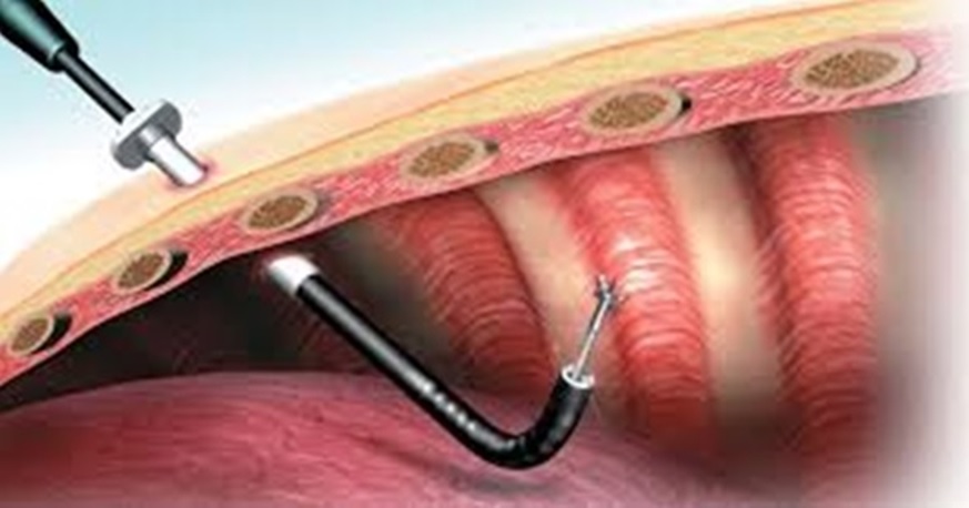
EBUS or Endobronchial Ultrasound is ultrasonic imaging of the mediastinal structures in the immediate vicinity of central airway and sampling the abnormal tissue using Fine Needle Aspiration process or FNA. Once lymph-nodes or masses are in central chest area called mediastinum sampling these lesions becomes difficult. Approaching these lesions thru air-way using Endobronchial ultrasound is very convenient.
Prior to this advancement patients had to undergo an extensive surgical process called Mediastinoscopy for evaluation of these lesions. Mediastinoscopy was a surgical process requiring general anesthesia, operation theater and had a higher risk.
EBUS is done in bronchoscopy suite under deep sedation. It’s a pain-less procedure and after sedation there is practically no discomfort to the patient. Sample obtained thru the procedure is aqequate enough to confirm the diagnosis, ascertain the sub types of cancer and even determine the genetic mutations in the cancer cells in most of the cases. Tuberculosis, Sarcoidosis, Lymphoma, Primary lung cancers and Metastatic mediastinal deposits or lung masses can be confirmed by samples obtained thru procedure.
Patient preparation is similar to a bronchoscopy, fasting of 5 hours prior to procedure is must. Procedure would last for 30 -60 mins depending upon number of stations to be sampled and the diagnosis under question. Post-procedure patient will be under observation for an hour and can have meal after 2-3hours. Procedure can be done in daycare , in special cases patient may need hospital admission.
Patient preparation:
Keep NPO ie nil orally for at least 5 hrs prior to procedure (no food/no liquids/no water)
Pre check- platelet count and coagulation profile.
Tell your doctor if you are diabetic and ask to avoid your diabetes drug
Blood thinners (like clopidogrel/ Aspirin) might need to be stopped , ask the doctor
Always mention if you have any specific allergies
You may take your regular medicine of blood pressure and thyroid(hypothyroidism) after discussing with doctor.
Thoracoscopy
Pleuroscopy or medical Thorascopy is the endoscopic procedure to peep into the pleural space. This is the space surrounding the lungs from outside. In normal scenario this is a small space containing little amount of lubricating fluid to avoid any friction between two layers covering the lungs during lungs movement of respiratory phases. In a large number of diseases there may be collection of huge amounts of abnormal fluid in this space. At times even lungs may rupture leading to collection of air around lungs. In both the cases a patient would need insertion of a tube to drain out abnormal collection of fluid/air outside lungs.
If the cause of fluid collection is not ascertained by initial investigations, in those scenarios’ biopsy of pleura the covering of lungs would be required. This biopsy, visualization and drainage of fluid is possible by procedure of Pleuroscopy/Medical thoracoscopy.
This is a minimally invasive procedure. After localizing fluid in the chest cavity using ultrasound study, site is marked and surrounding skin is anaesthetized with local anesthetic agent. Intravenous pain killers are given as per the need. Chest trocar is inserted pleural endoscope is advanced through chest cannula . PLaural surfaces can be visualized after draining the fluid, Pleural biopsy is obtained from outer covering (Parietal Pleura). This sample is sufficient for microbiological study, Cytology, histopathology, Immuno- Histo-chemistry (IHC) and genetic mutations.
Post-procedure a draining tube is usually left in the place to drain remaining collection until the lung inflates fully .